Microscope Images
Fiber Images in Natural Light Under Microscope
Used Fibers
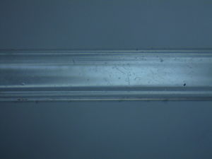
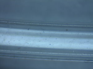
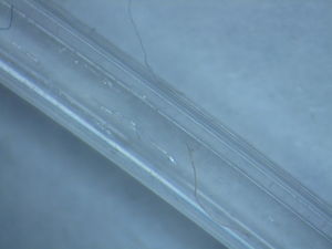
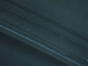
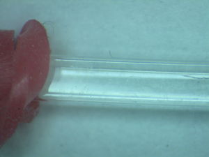
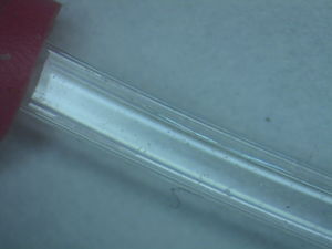
Presented below are images of a fiber segment that had been processed and bent according to procedures discontinued or updated by January 2015. These procedures included cleaning with solvents, temporarily bundling with velcro straps, labeling with sticky tags, and straightening and bending with heat and hot water. Additionally, these fibers may have been exposed to glue and/or paint. In the time between their handling and these images, they were handled as scraps; therefore, it is hard, if not impossible, to attribute their damage to previous handling procedures. However, examining their scratches and markings provides insight into their susceptibility to damage.
Figure 1 exhibits dust from the environment and rubber from the researcher's gloves. There appears to be a slight film over the waveguide, which might be from a combination of normal dust and humidity.
